The Role of Virtual Reality in Cell Microscopy
dr. R. van Liere
Centrum voor Wiskunde en Informatica
|
Confocal and deconvolution microscopes allow biologists and
biomedical researchers to obtain time dependent 3D data sets of
biological objects, such as cells and tissues. Scientific
visualization can provide effective visual presentations of structural
characteristics of these data sets. This talk addresses the role of
virtual reality in gaining insight in these presentations. The
argument is made that the understanding of structural characteristics
of time dependent 3D confocal cell data requires spatial judgments.
Perceiving these characteristics can best be gained using virtual
reality device technology.
I will discuss our experiences in working with cell biologists and discuss the pros and cons of using virtual reality for the analysis of 4D cell data. We will sketch the Proteus system, a virtual environment we have developed for studying 4D confocal cell data. This system Proteus is being been used on a large fish tank display with a 6 DOF wand for interaction. I wil discuss the pros and cons of using Proteus in other environments, such as the CAVE, HMDs, desktop workstations and with different input devices. |
|
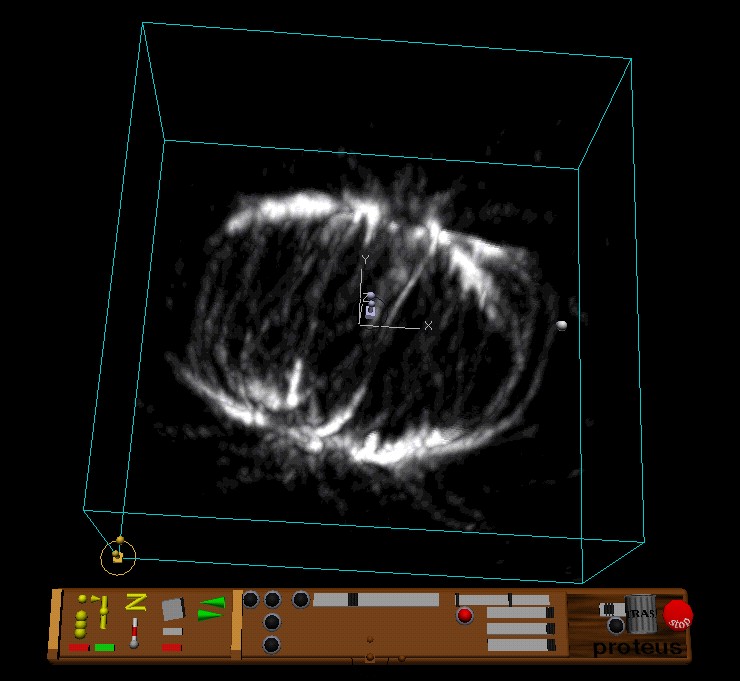
| An example of a spindle structure from a biological cell and the Proteus interface elements used to control the view on this object |
Lezing, te presenteren op het symposium 'Echt of Namaak: simulatie en modellering in beweging', 5 oktober 2001, Rijksuniversiteit Groningen.